The second article was printed in the journal of the american dental association, and discusses the effectiveness of barrier sheaths. Dental x ray film has the following characteristics:
After the film has developed in a short time like a few minutes, your dentist can determine what needs to be done and the method of treatment.

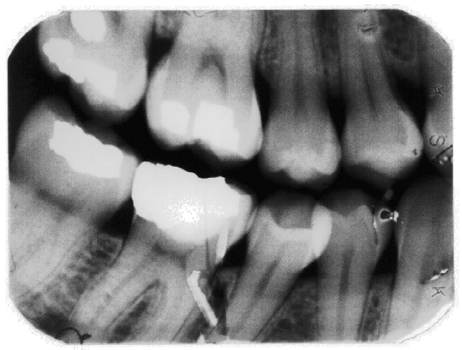
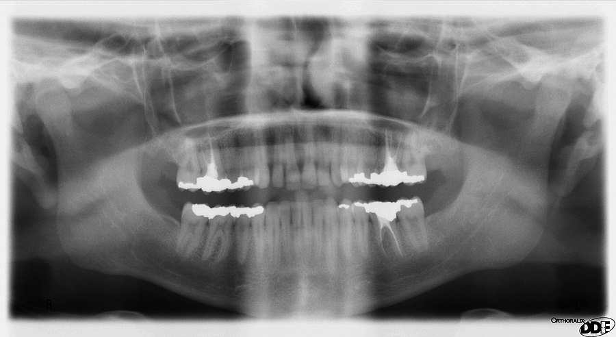
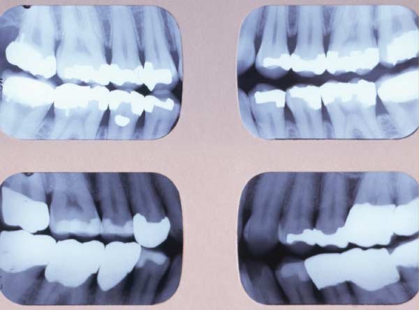
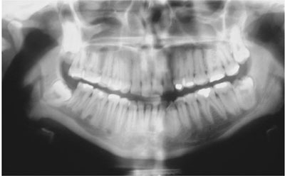
How to read dental x-ray film. Dental radiology “cheat sheet” area imaged general technique and tips lower pm and m place film in vestibule between the tongue and teeth. The green arrows are pointing to the bone. A small film is placed inside the mouth next to the tooth.
Periapical films, since often, there is not enough room in the mouth to place film parallel to the teeth. Dental x ray film has the following characteristics: Ezdicom — ( windows) this software.
Structures that are dense (such as silver fillings or metal restoration) will block most of the. Dentists use radiographs for many reasons: After the film has developed in a short time like a few minutes, your dentist can determine what needs to be done and the method of treatment.
Due to this, hard tissues like the enamel and dentin will appear light in color. The blue arrows are pointing to the healthy enamel. The dentin layer is between the enamel and the pulp.
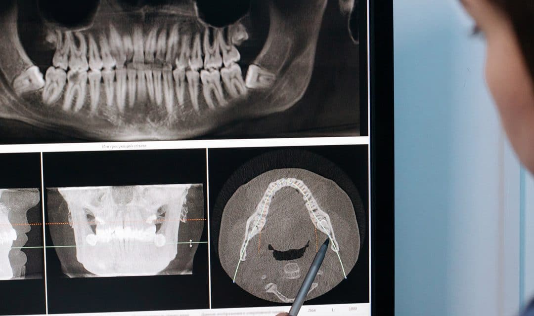
Dental Imaging Explained – Orthodontic Associates

Dental X-rays How To Tell If You Have A Cavity Fridley Mn
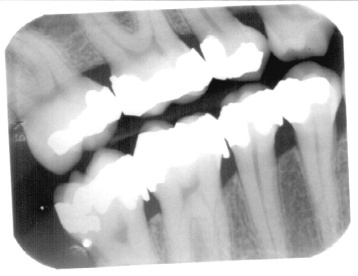
Scanning X-ray Films Dealing With The X-ray Films
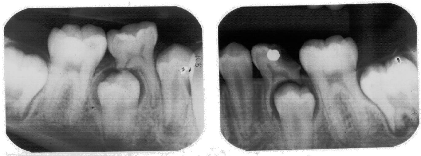
Scanning X-ray Films Dealing With The X-ray Films
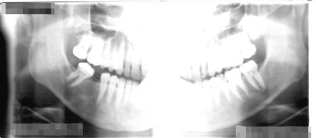
Scanning X-ray Films Dealing With The X-ray Films
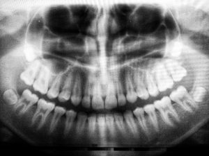
Understanding Dental X-rays – Dawson Dental
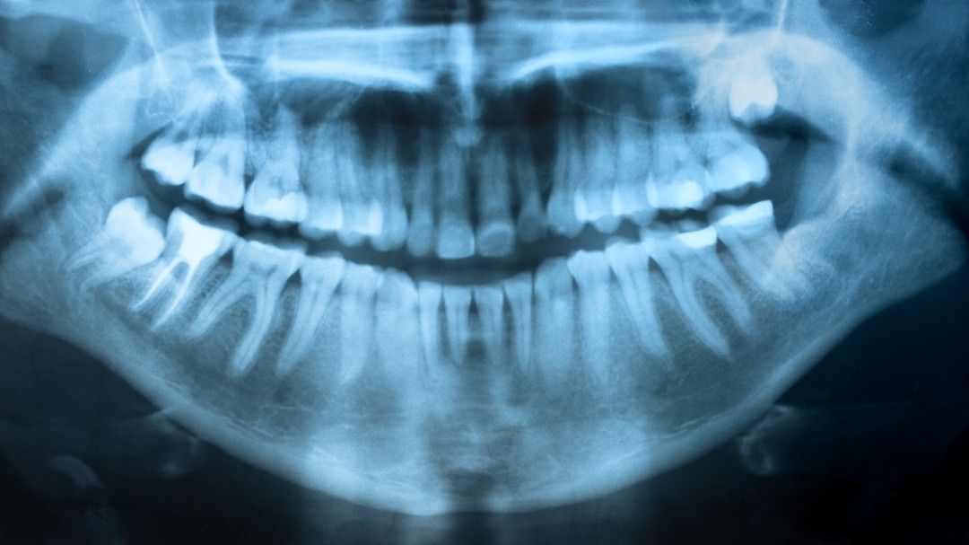
How Safe Are Dental X-rays – North Sydney Dental Practice

Components Of X Ray Film Packet – Youtube

Image Evaluation For An Fmx- Identifying Correcting Errors – Youtube

Dental X-rays – Dental Health Today Dental Health Today
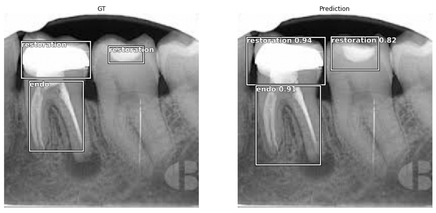
Fastai Object Detection Applied To Dental Periapical X-rays By John Persson Analytics Vidhya Medium


